CT Scans in Lung Cancer Detection: An In-Depth Review


Intro
Lung cancer remains one of the most formidable challenges in oncology today. The insidious nature of this disease often means it goes undetected until it reaches advanced stages, when treatment options become limited. In this context, the role of advanced imaging techniques, specifically CT scans, is of paramount importance. This article ventures into the depths of how CT scans enhance the early detection of lung cancer, a necessity that could save countless lives.
Research Context
Background Information
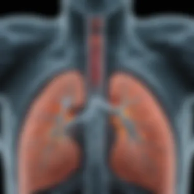
The evolution of lung cancer diagnosis has taken significant leaps since the early days of chest X-rays. CT imaging, due to its intricate cross-sectional views of the lungs, offers precision in identifying nodules and masses that may be invisible on standard imaging modalities. A large part of lung cancer screening programs today incorporate low-dose computed tomography to seek out early signs of malignancy in high-risk populations.
Importance of the Study
The necessity of understanding the capabilities and limitations of CT scans is not just an academic exercise; it holds real-world implications for patient outcomes. By dissecting the modalities used in lung cancer detection, this study underscores why early diagnosis via CT imaging could alter the course of the disease. Furthermore, examining diverse patient populations aids in identifying potential disparities in lung cancer detection and treatment, which is crucial for improving healthcare equity.
Discussion
Interpretation of Results
Results surrounding CT scan efficacy paint a promising picture. Studies indicate that low-dose CT scans can reduce lung cancer mortality by 20% compared to traditional X-rays. Patients who undergo regular screenings are more likely to catch the disease before it spreads. This sharp contrast in outcomes, however, comes with its own set of worries regarding overdiagnosis and the accompanying healthcare costs.
Comparison with Previous Research
Historically, lung cancer was often diagnosed too late due to the limitations of older imaging techniques. Previous research typically highlighted the role of X-rays, which fell short in sensitivity. Modern studies underscore the transformative effect CT scans have on this landscape. A meta-analysis shows a consistent trend where CT imaging identifies more lung cancer cases, reinforcing the argument for implementing CT scanning in routine screenings for high-risk individuals.
"The difference in detection rates between traditional X-rays and CT scans is like comparing apples and oranges. CT scans simply yield more productive results when it comes to early diagnosis of lung cancer."
By examining these studies, we gather insights that not only validate the current methodologies used in lung cancer screening but also illuminate the path forward as advancements continue to unfold. Ultimately, it is this rich tapestry of research and clinical application that fortifies the crucial role of CT scans in modern oncology.
Prologue to Lung Cancer
Lung cancer remains one of the most formidable challenges in the realm of oncology. This section lays the groundwork for understanding this dire disease, underlining the significance of examining how CT scans can assist in its detection. The importance of diving deep into this subject cannot be overstated, as early detection can lead to more effective treatment options and ultimately save lives.
In discussing lung cancer, it is crucial to contextualize its prevalence and impact. Lung cancer, primarily caused by smoking, environmental factors, and genetics, manifests through insidious symptoms often mistaken for less serious ailments. By honing in on early signs and symptoms, healthcare professionals can strategize better detection methods, paving the way for improved patient care.
Understanding Lung Cancer
Lung cancer is primarily classified into two main types: non-small cell lung cancer (NSCLC) and small cell lung cancer (SCLC). NSCLC is more prevalent, accounting for about 80% of cases, and generally has a slower progression compared to its small cell counterpart, which tends to be more aggressive.
Diagnosing lung cancer often begins with an evaluation of the patient’s history, physical exams, and imaging tests. Risk factors such as age, smoking habits, and exposure to carcinogens can provide valuable insight for clinicians. Further investigations, like bronchoscopy or biopsies, are then performed to confirm diagnosis and stage the cancer.
"Understanding lung cancer is not just about recognizing symptoms but also about grasping the broader environmental and hereditary landscape that contributes to its development."
Statistics and Prevalence
The statistics surrounding lung cancer paint a stark picture. According to the American Cancer Society, nearly 240,000 new cases of lung cancer are expected annually in the United States alone. Globally, lung cancer is the leading cause of cancer-related deaths, surpassing breast, colorectal, and prostate cancers combined.
Considering these numbers, it's imperative to acknowledge the varying incidence rates among demographics. Contributing factors that manifest in different populations include socioeconomic status, access to healthcare, and lifestyle choices. Understanding these trends not only highlights disparities in lung cancer treatment but also emphasizes necessary public health interventions.
Key Statistics:
- Incidence: Lung cancer accounts for about 13% of all new cancer diagnoses.
- Mortality: It leads to more deaths each year than breast, prostate, and colorectal cancers combined.
- 5-Year Survival Rate: The average 5-year survival rate stands around 18%, but with early detection, this can rise significantly.
In summary, the urgency in addressing lung cancer cannot be ignored. As this article unfolds, we will explore how CT scans play a pivotal role in detecting lung cancer early, contributing significantly to survival outcomes.
The Significance of Early Detection
Early detection of lung cancer plays a pivotal role in improving patient outcomes. The earlier the disease is identified, the better the chances of successful treatment and long-term survival. Many lung cancer cases go unnoticed until they reach an advanced stage, where interventions become limited and outcomes grim. Thus, increasing awareness about the importance of early detection is critical.
One of the primary benefits of early detection is the substantial increase in survival rates of lung cancer patients. Understanding the cancer's stage at diagnosis can be a game-changer. When diagnosed at a localized stage, the five-year survival rate can soar above 55%, compared to less than 5% for cases detected at a distant stage. This stark contrast underlines how essential it is to catch lung cancer early on.
However, the path to early detection is fraught with challenges. Many individuals may not experience noticeable symptoms until the disease has progressed. This delay often comes from the insidious nature of lung carcinomas, coupled with common symptoms that could be attributed to other less dangerous conditions, such as chronic bronchitis or asthma. Another issue stems from the fact that screening guidelines may not be effectively communicated or adhered to, leaving at-risk populations with limited access to potentially life-saving assessments.
"The key to better outcomes lies not just in technology, but also in understanding and navigating barriers to access and awareness for at-risk populations."
In addition, there are psychological, social, and financial barriers that can hinder early diagnosis. Many individuals may fear the repercussions of a positive diagnosis, avoiding screening altogether. There are also groups within the population, such as the uninsured or those in rural areas, who might lack access to advanced imaging technologies like CT scans. This inequity complicates the landscape of lung cancer screening and detection.
Thus, while the narrative surrounding early detection is one of hope and improvement, it must also acknowledge the nuanced realities that patients face. Recognizing that the quick identification of lung cancer significantly impacts survival rates sheds light on the numerous factors influencing early diagnosis. Engaging in proactive conversations about these issues can spur collective action toward achieving better patient outcomes.
CT Scans: An Overview
In the realm of modern medicine, CT scans hold a prominent place, especially in the detection of lung cancer. Their role is not just significant; it's pivotal. A CT scan, or computed tomography scan, combines a series of X-ray images taken from multiple angles and uses computer processing to create cross-sectional images of bones, blood vessels, and soft tissues inside the body. In terms of lung cancer, the clarity of these images can be a game changer, offering insights that other imaging techniques may miss.
What is a CT Scan?
A CT scan is like a finely-tuned, high-tech camera that captures detailed images of the body from different angles. Unlike a regular X-ray, which produces a flat image, CT scans create a detailed 3D representation of internal structures. This precision is crucial for detecting anomalies, such as tumors or lesions in the lungs.
For many patients, the word "scan" might evoke apprehension. However, the process is generally quick and straightforward. Patients lie on a table that moves through a large, doughnut-shaped machine. As this happens, the scanner takes numerous images of the area of interest, which the computer assembles into a comprehensive visual representation.


In lung cancer detection, these images can reveal very small nodules, which may signal the early stages of cancer. The technology focuses not solely on size but also on the characteristics of these nodules, distinguishing benign from malignant growths effectively.
How CT Scans Work
So, how does this technology work its magic? CT scans utilize X-ray technology in tandem with advanced computer algorithms. When a patient is scanned, the machine emits a beam of X-rays that passes through the body. Denser structures like bones absorb more radiation, appearing lighter in the resulting images. Conversely, softer tissues, such as those in the lungs, allow more X-rays to pass through.
- Image Acquisition: After the X-ray source and sensor rotate around the patient, they continuously collect data. At this stage, the patient is often instructed to hold their breath to minimize movement and blur in the images.
- Data Processing: The collected data is sent to a computer, which reconstructs the images using complex algorithms that account for various factors, including the angles and intensity of X-ray beams.
- Visual Output: The end result is a series of cross-sectional images that healthcare professionals can analyze from different perspectives.
Interestingly, advancements in CT technology have led to quicker scans with improved resolution. Enhanced lung imaging techniques can even allow for the detection of nodules as small as 1-2 mm, a size that would previously have been too tiny for reliable identification.
It’s clear that the CT scan is not just a routine procedure but a critical tool that helps in the early detection and staging of lung cancer. As technology continues to advance, the potential for these scans to enhance the diagnostic process will only improve, promising better outcomes for patients.
Comparative Imaging Techniques
Understanding imaging techniques is vital in today's clinical landscape, especially when it comes to diagnosing lung cancer. Each imaging modality offers its own set of benefits and limitations. In this segment, we will closely examine how CT scans stack up against other imaging forms such as X-rays, MRIs, and PET scans. By doing so, healthcare providers can make informed decisions tailored to individual patient needs, ultimately leading to improved outcomes.
CT Scans vs. X-rays
CT scans and X-rays serve different purposes in medical evaluation. While X-rays can provide a quick look at the lungs, they often lack the detail necessary for a definitive lung cancer diagnosis. For instance, in a case of lung nodules, a simple X-ray may not reveal subtle changes that a CT scan would capture.
- Advantages of CT Scans:
- Limitations of X-rays:
- High-resolution images reveal small lesions, which increase the chances of early detection.
- Multi-dimensional views allow for a thorough examination of surrounding tissues, improving diagnostic accuracy.
- Generally ineffective for identifying small lesions that could signify malignancy.
- Two-dimensional images can obscure overlapping structures.
Ultimately, the distinction between CT scans and X-rays becomes a crucial part of the initial evaluation process and affects subsequent treatment steps.
CT Scans vs. MRI
While CT scans excel in identifying lung cancer, MRIs also offer valuable insights, particularly for assessing soft tissue characteristics. This technique uses magnetic fields and radio waves, giving it a unique edge in certain scenarios. However, this does not make it superior for lung imaging.
- Benefits of Using MRI:
- CT Scan Strengths:
- Excellent for soft-tissue differentiation, which is beneficial when examining adjacent structures.
- No radiation exposure, making it a preferable choice for some patients.
- More effective for lung imaging due to the ability to provide cross-sectional views that demonstrate how lesions interact with lung structures.
- Quick in comparison; CT scans typically take less time than MRIs, making it potentially less burdensome for patients.
In many cases, the choice between CT and MRI depends on the clinical question at hand, and understanding the strengths and weaknesses of each method is essential.
CT Scans vs. PET Scans
PET scans offer a completely different perspective, focusing primarily on metabolic activity instead of structural analysis. This differentiation is important when it comes to staging cancer and planning treatment. However, it’s important to recognize the roles they play together.
- Insights from PET Scans:
- Position of CT in Lung Cancer Detection:
- Provide functional information about how tissues are behaving, hence assisting in differentiating benign from malignant lesions.
- Can help in identifying metastasis that may not be evident in a CT scan.
- Offers detailed anatomical data, which is crucial for determining tumor size, location, and involvement with surrounding structures.
- High sensitivity in detecting early-stage lung cancer lesions that require immediate intervention.
In summary, CT scans, X-rays, MRIs, and PET scans have unique and complementary roles in lung cancer detection. Understanding how they interact can help in devising an effective detection and treatment strategy.
Ultimately, the choice of imaging technique will depend on multiple factors including patient history, symptoms, and initial findings.
Effectiveness of CT Scans in Lung Cancer Detection
The effectiveness of CT scans in lung cancer detection is a critical aspect of modern medical imaging. As lung cancer remains one of the leading causes of cancer-related mortality worldwide, it is vital for healthcare professionals to have accurate and reliable tools for early diagnosis. Here, we delve into the nuances of CT scan effectiveness, emphasizing its sensitivity and specificity, and the limitations inherent in this imaging technique.
Sensitivity and Specificity
When discussing the effectiveness of CT scans, two critical statistical measures come into play: sensitivity and specificity. Sensitivity refers to the ability of a test to correctly identify patients with a disease. A highly sensitive test will have fewer false negatives, meaning it will successfully catch most cases of lung cancer that exist.
In terms of lung cancer detection, low-dose CT scans stand out, boasting a sensitivity that is notably higher when compared to traditional chest X-rays. Research indicates that low-dose CT can detect lung cancers up to 92% of the time, a remarkable statistic when weighed against the approximately 70% sensitivity found in X-rays.
On the other hand, specificity reflects how well the test identifies those without the disease, which is equally significant. A high specificity means fewer false positives, crucial for minimizing unnecessary anxiety and invasive procedures for patients. While CT scans can efficiently identify lung cancer, they sometimes struggle with specificity due to overdiagnosis—detecting nodules that may not pose any real threat. In clinical practice, it’s essential for radiologists to interpret these results judiciously, discerning between actionable findings and incidentalomas that may never cause harm.
Statistical insights illustrate the balance that CT scans must strike between sensitivity and specificity.\n- Low-Dose CT Sensitivity: Approaches 92%
- Traditional X-ray Sensitivity: Approximately 70%
- CT Specificity: Can vary, often lower than sensitivity, warranting careful analysis.
Limitations of CT Imaging
Despite the power of CT scans as a diagnostic tool, they are not without their limitations, which need to be thoroughly considered.
- Radiation Exposure: One significant drawback is the radiation involved in CT imaging. Although the levels are relatively low, repeated exposure can cumulatively raise the risk of developing cancer later in life, a concern particularly in younger patients. This aspect calls for a careful evaluation of risks versus rewards in screening programs.
- Interpretation Variability: The accuracy of CT scans heavily relies on the skill of the radiologist interpreting the images. Variability in interpretations can lead to misdiagnosis or delayed detection, emphasizing the need for continued education and standardized protocols in imaging centers.
- Overdiagnosis: It’s crucial to recognize that not all detected tumors require treatment. The phenomenon of overdiagnosis can lead patients to undergo unnecessary interventions, causing more harm than good. Therefore, a clear dialogue about potential false positives and the nature of certain findings is essential.
"The balance of effectiveness lies not just in detection, but in judicious management of indeterminate findings."
- Cost Factors: Lastly, the financial burden associated with CT scans can be a limiting factor. They are more expensive than other imaging techniques, which may pose accessibility issues in under-resourced settings.
Screening Guidelines and Recommendations


Screening for lung cancer is a complex and significant aspect of public health strategy aimed at early detection, which can certainly lead to better prognoses. Screening guidelines aim to identify individuals who are most likely to benefit from lung cancer screening while minimizing potential harms. These guidelines are informed by extensive research and analysis, highlighting the necessity for tailored recommendations that consider both the risks of lung cancer and the effectiveness of CT scans in detecting tumors at an earlier, more treatable stage.
Who Should Get Screened?
When discussing who should undergo screening for lung cancer, understanding the risk factors becomes crucial. The current guidelines primarily advocate for high-risk groups, particularly:
- Long-term smokers: Individuals aged 50 to 80 who have a history of heavy smoking. Specifically, those who have smoked a pack a day for 20 years or two packs a day for 10 years.
- Former smokers: People who quit smoking less than 15 years ago fall into this category as well.
- Occupational exposure: Those exposed to asbestos or other carcinogens in the workplace are also at heightened risk.
In light of growing evidence, it's vital to engage in proactive discussions about other factors such as family history of lung cancer and pre-existing lung diseases like COPD. Often overlooked, these elements can influence the necessity for screening and should be factored into individualized assessments.
Frequency and Protocols
The frequency of screenings isn’t a one-size-fits-all scenario. Generally, the recommendation is an annual low-dose CT scan for individuals in the high-risk groups identified earlier. Adherence to strict protocols is essential in ensuring that these screenings are effective and do not lead to unnecessary anxiety or additional medical procedures. Key points in the protocols include:
- Initial Screening: Yearly low-dose CT scans are suggested to establish a baseline and monitor any changes over time.
- Follow-ups: Depending on the findings, follow-up protocols may vary widely. For instance, if a nodule is detected, further imaging or biopsies may be recommended based on size and characteristics.
- Risk Assessment Tools: Utilizing validated tools to assess risk periodically can also enhance screening practices, ensuring that only those who genuinely need it stay in the loop.
Advancements in CT Scan Technology
The landscape of medical imaging has transformed significantly over the years, and the advancements in CT scan technology stand at the forefront of these changes. As lung cancer detection becomes increasingly vital, these improvements ensure a more accurate, efficient, and patient-friendly approach to diagnostics. This section delves into the emerging technologies and innovations that are setting the stage for better lung cancer detection and management.
Emerging Technologies
Current advancements are nothing short of revolutionary. For one, high-resolution CT scans enhance the clarity of images, allowing radiologists to discern even the faintest signs of lung pathology. Unlike older models, these modern iterations can detect smaller nodules that might have been previously missed, effectively increasing early detection rates of lung cancer.
- Low-dose CT scanning is another breakthrough, which typically reduces radiation exposure without sacrificing image quality. This is particularly important for screening purposes, as it enhances patient safety while providing crucial diagnostic insights.
- Dual-energy CT technology further enriches the capability to differentiate between various types of tissues and fluids within the lungs. This not only aids in identifying tumors but also helps in assessing their characteristics more accurately than plain imaging techniques.
- AI-enhanced algorithms are also sparking interest, capable of identifying anomalies in scans with impressive accuracy. This fusion of artificial intelligence with CT technology often leads to faster interpretations, expediting decision-making for patient care.
"The blend of artificial intelligence with traditional imaging methods could redefine diagnostic accuracy for lung cancer, presenting a clearer picture of disease development and treatment options."
Innovations in Imaging Techniques
Innovations in CT scan technology extend beyond just hardware improvements. Software enhancements are key players in refining imaging techniques. For example, image reconstruction algorithms now utilize sophisticated computing methods to produce images that highlight abnormalities in the lungs while minimizing artifacts. This ensures that the results are not only clearer but also more confident in diagnosis.
In addition, motion correction technologies are being developed to counteract the effects of patient movement during scans. This is paramount for achieving consistent image quality, particularly in patients who may have difficulty staying still. By reducing blurring and distortions, these innovations enhance the diagnostic utility of CT scans.
Clinicians and researchers are also looking into functional imaging methods, which assess not just the structural but also the physiological aspects of the lungs. Techniques like CT perfusion imaging can provide insights into blood supply and overall lung function, offering a broader perspective on how lung cancers might interact with surrounding environment and tissues.
These advancements collectively underscore a critical shift in how lung cancer is diagnosed and monitored. As technology continues to push boundaries, the potential for improved patient outcomes through better imaging grows exponentially. It renders a clear awareness that the future of lung cancer detection is not just dependant on better technologies, but also on their integration into clinical practice to foster a supportive environment for effective patient care.
Interpreting CT Scan Results
Understanding the outcomes of a CT scan is a critical step in the process of diagnosing lung cancer. These scans generate detailed images of the lungs, enabling radiologists and medical professionals to assess for the presence of tumors, nodules, or other anomalies that might suggest malignancy. Accurate interpretation of these results can significantly impact treatment decisions and, ultimately, patient outcomes.
When a patient undergoes a CT scan for lung cancer screening, there are several key elements that specialists must consider. First and foremost, they need to recognize the appearance of different types of lung lesions. For instance, benign nodules often have smooth, round edges, while malignant tumors may appear irregular with spiculated borders.
Moreover, understanding the size and growth rate of detected nodules plays a pivotal role in diagnosis. Studies show that nodules larger than 8 mm warrant closer observation. If these nodules double in size within a short period, this often raises red flags for lung cancer.
In summary, diving deep into the specifics of CT scan outcomes is essential for timely intervention. The insights gleaned from these interpretations form the backbone of early cancer detection strategies.
"Timely interpretation of CT scans can be the difference between successful treatment and missed opportunities."
Understanding Scan Outcomes
Navigating the complexities of CT scan results is no small feat. Radiologists need to grasp not only the clear positives or negatives but also the shades of gray in between. Key points to be mindful of include:
- Nodule characteristics: Size, shape, and surface characteristics can help distinguish benign from malignant.
- Location of the findings: Certain areas of the lungs are more prone to malignancies and can guide subsequent evaluations.
- Presence of lymph nodes: Enlarged lymph nodes might indicate metastasis, which changes the stage of the cancer.
Each of these factors can contribute to a more nuanced understanding of the patient's condition and aid in directing further diagnostic efforts, such as biopsies or additional imaging.
Role of Radiologists
The radiologist's role in interpreting CT scan results is multifaceted and critical in the context of lung cancer detection. These specialists are not just technicians; they are detectives unearthing vital clues hidden within the images. Their expertise is essential in differentiating between benign conditions, such as infections or scarring, and potential tumors.
Radiologists must communicate findings clearly to other healthcare providers. They often provide detailed reports that outline not just what was seen but also convey the urgency of follow-up actions. Here are some aspects of their responsibilities:
- Assessment of imaging features: Radiologists must analyze every element of the scan, documenting their observations with precision.
- Collaboration with oncologists: Radiologists frequently work alongside oncologists and primary care physicians to determine the best course of action based on imaging findings.
- Continual education: Given the rapidly evolving nature of imaging technologies and cancer diagnostics, radiologists engage in ongoing education to stay updated.
In essence, the role of the radiologist is pivotal in bridging the gap between imaging technology and clinical care.
Case Studies in Lung Cancer Detection
Case studies in lung cancer detection are more than mere anecdotes; they serve as invaluable teaching tools, showcasing the real-world implications of both effective and ineffective diagnostic practices. The importance of this topic lies in the insights gleaned from individual patient experiences, which can illuminate broader trends and highlight beneficial practices in early diagnosis and management. With each case, the intricate dance between technology and human interpretation becomes apparent, offering a glimpse into how CT scans play a pivotal role in identifying lung cancer at various stages.
Successful Early Diagnoses
Successful early diagnoses often hinge on the precise interpretation of CT scans. Take, for example, a 62-year-old man, a long-time smoker, who attended his annual health check-up. A routine low-dose CT scan revealed a small nodule in his right lung. Rather than dismissing this finding, the medical team opted for a follow-up CT scan after three months. This cautious approach paid dividends; the nodule, initially 3 mm in diameter, had increased in size and exhibited worrying characteristics indicative of malignancy. Prompt biopsies confirmed stage I lung cancer, allowing timely intervention and a targeted surgical resection. This case underscores how CT scans can reveal abnormalities that, if unaddressed, could transition from being manageable to life-threatening.
Complex Cases and Challenges


While some cases exhibit stark clarity, others are cloaked in complexity. Consider a 55-year-old woman with a family history of lung cancer, presenting with chronic cough and unexplained weight loss. Initial CT scans appeared unremarkable, but she continued experiencing symptoms. The challenge here lay in balancing vigilance against the risks of overdiagnosis. After multiple consultations, a second opinion led to more advanced imaging techniques. Further investigation revealed a hidden malignancy that the first scans failed to detect. This case demonstrates that CT scans, while critical, may not always suffice alone. It also speaks volumes about the necessity of comprehensive diagnostic protocols, which should include detailed patient history and, if required, supplementary examinations.
"In every case, while technology provides us the tools to detect, it's our clinical judgment that ultimately shapes outcomes."
The lessons drawn from these cases highlight the intricate interplay of technology and clinical decision-making in lung cancer detection. Successful early diagnoses can save lives, but complex cases remind us that vigilance and a thorough diagnostic approach are essential to meet the realities of each individual patient's circumstances. It’s crucial that the medical community continues to learn from these case studies to improve patient outcomes and enhance our understanding of lung cancer detection.
Ethical Considerations in CT Screening
The realm of medical imaging isn't solely grounded in advancements in technology and techniques; it grapples with equally pressing ethical realities that impact patient care, trust, and public health. In lung cancer detection, the use of CT scans demands a careful balancing act of providing critical information while respecting patient autonomy. The stakes are high, as the implications of screening extend beyond mere diagnostics.
When discussing ethical considerations in CT screening, several core elements emerge: informed consent, risk versus benefit analysis, and the broader implications for healthcare equity. Thus, it becomes clear that ethical scrutiny is not a peripheral concern; rather, it is integral to fostering a responsible framework for patient engagement, ensuring that the benefits of early detection do not come at the cost of undue stress or harm.
Informed Consent
Informed consent stands as a cornerstone of ethical medical practices. At its essence, it refers to the process through which patients are educated about the procedures they will undergo, including the risks, benefits, and alternatives. For CT screenings, patients must fully grasp what the scan entails.
This includes understanding the nature of radiation exposure, which, despite being relatively low, might still provoke anxiety in some individuals. Patients should be apprised of the possibility that a CT scan could lead to false positives, resulting in unnecessary follow-up tests or invasive procedures. Conversely, patients also need to be informed about the potential for missed cancers or lesions that may not be detectable via CT imaging.
The challenge lies in effectively communicating these complexities. A one-size-fits-all approach won't suffice; healthcare professionals must tailor their explanations to meet individual patients' needs and comprehension levels. This means using clear, straightforward language rather than medical jargon. For instance, when discussing follow-up procedures, a doctor might say,
"If the scan shows something unusual, we might need to look closer with another type of test or even a biopsy. It's like checking your mailbox for a letter; just because you found one does not mean you need to worry just yet."
This kind of analogy can help demystify the process.
Risk vs. Benefit Analysis
When weighing the ethical dimensions of CT screening, the risk versus benefit analysis is paramount. On one hand, the potential for early lung cancer detection through CT scans can significantly enhance survival rates. Hospitals and clinics have witnessed firsthand how timely intervention can drastically change outcomes for patients.
However, benefits must be examined in light of the associated risks. Unintended consequences, such as overdiagnosis, might lead patients down an unnecessary path of anxiety and treatment for slow-growing tumors that might never cause harm. Such situations raise valid questions about whether the distress caused by screening outweighs the actual health benefits derived from interventions.
Healthcare providers are tasked with navigating these murky waters. They must engage in frank discussions with patients about their individual risk factors for lung cancer, your smoking history, family medical background, and lifestyle choices. This personalized approach ensures that patients make informed decisions based on their specific context, balancing the immediate benefits of detecting potential health threats against the psychological and physical costs of unnecessary interventions.
In essence, both informed consent and risk versus benefit discussions engender a landscape of transparency in healthcare that empowers patients while guiding them through complex choices. As CT technology advances, this ethical lens becomes crucial in stewarding responsible care that prioritizes the well-being of individuals while leveraging innovative diagnostic tools.
Future Directions in Lung Cancer Imaging
As we look ahead, the landscape of lung cancer imaging is poised for significant transformations. The integration of advanced technologies promises not just improvements in how we detect lung cancer but also how we interpret and act on those findings. Advances in imaging techniques will likely influence patient outcomes, streamline workflows in clinical settings, and present new avenues for personalized treatment plans. Here, we explore two specific avenues of progress: the integration of artificial intelligence (AI) and the potential for enhancements in early detection capabilities.
Integrating AI with Imaging
The rise of artificial intelligence signals a new chapter in medical imaging. AI algorithms are being trained to analyze vast datasets quickly and with unprecedented precision. This capability not only assists radiologists in identifying anomalies but potentially reduces the risk of human error in readings. For instance, AI can be programmed to recognize subtle patterns in lung scans that might be missed by the naked eye.
- Enhanced Accuracy: Machine learning models can be refined over time, learning from previous scans to refine their predictive capabilities. This means that, over time, these tools may outperform even experienced radiologists in some aspects of analysis.
- Efficiency in Workflow: With AI taking on initial assessments of scans, medical professionals can allocate more time to complex cases requiring nuanced interpretation, ultimately leading to faster diagnoses for patients.
"The fusion of technology with traditional methodologies brings about redefine standards of care in lung cancer detection."
Potential for Early Detection Enhancements
Early detection remains a cornerstone in the fight against lung cancer. As research progresses, there are several potential enhancements that could revolutionize how we identify lung pathologies at their most treatable stages.
- Biomarker Integration: Exploring the combination of imaging techniques with biomarker testing could create a more holistic approach. For example, a CT scan could be used alongside blood tests that identify specific proteins or genetic material linked to lung cancer.
- Improved Screening Protocols: Tailoring screening protocols based on individual risks and genetic predispositions could lead to more targeted imaging strategies. This shift away from a one-size-fits-all could elevate early detection rates in high-risk populations significantly.
- Dynamic Imaging Techniques: As technology advances, we might see the emergence of dynamic imaging methods that allow for real-time observation of lung changes. Such techniques might enhance our understanding of tumor growth patterns, ultimately informing more effective interventions.
Patient Perspectives on CT Scans
When diving into the complexities of diagnosing lung cancer, it's crucial to consider the perspectives and experiences of the patients undergoing CT scans. Understanding how patients view and experience the process can shed light on its significance and the outcomes of detection methodologies.
Many patients encounter CT scans as a routine part of the diagnostic process, but the emotional and psychological aspects are often overlooked. Beyond just the technical procedure, the anxiety stemming from uncertainty about health can be overwhelming. Patients might describe the waiting period for results as the "waiting game," where every tick of the clock feels like an eternity. This emotional backlog can largely influence their perception of how effective and essential these scans are.
Experiences and Insights
Patients frequently share insights that punctuate the importance of CT scans in their journeys. For those with a lung cancer diagnosis, the clarity offered by these scans can be a double-edged sword. On one hand, there's relief in having a precise understanding of their condition. Conversely, the knowledge of having a diagnosis can also introduce fear of the unknown. Patients have expressed that hearing their doctors explain images showing potential malignancies can feel like a punch to the gut. They might say, "It’s like standing on the edge of a cliff, peering into the abyss."
Many recount feeling a degree of control regained when they receive detailed information from health professionals. This transparency allows patients to participate actively in their treatment plans, fostering a sense of empowerment over their choices. The trust relationship formed during this process can be paramount, as patients are more inclined to share all their concerns when they feel informed.
"It's not just about the scan; it's about how the results shape my life, my choices."
Barriers to Accessing Care
Despite the significant value of CT scans, numerous barriers can impede access to these critical diagnostic tools. Various factors contribute to these challenges, affecting patient experiences and outcomes. Financial constraints can weigh heavily on individuals and families. For instance, some insurance plans may limit coverage or impose high out-of-pocket costs for lung cancer screening, forcing patients to weigh their options carefully. In areas with limited healthcare infrastructure, finding facilities equipped for advanced imaging can become a daunting task.
Additionally, geographical disparities result in uneven access to care. Patients living in rural or underserved urban areas might find themselves traveling long distances to receive the necessary scans. The transport burden can strain both their time and financial resources, often leading to delayed diagnoses and treatment.
Cultural factors also come into play. Some patients may have reservations or preconceived notions about medical imaging, stemming from historical mistrust in healthcare systems. Language barriers and lack of information regarding procedures can further alienate some demographic groups, reinforcing inequality in accessing lifesaving technology.
In summary, integrating patient perspectives into the discourse about CT scans highlights the multifaceted nature of lung cancer detection. Understanding their experiences and the barriers they face tailors a more comprehensive narrative around these diagnostic tools, emphasizing the urgent need for improved accessibility and support.
Endings on CT Scans and Lung Cancer Detection
In piecing together the insights from this comprehensive examination, it becomes quite clear that CT scans hold a crucial position in the detection of lung cancer. The wealth of knowledge accrued throughout the article underscores not only the technicalities of how CT imaging operates but also its tangible impact on patient outcomes. In an age where early detection can often be the difference between life and death, the role of CT scans cannot be overstated. Their ability to illuminate the intricate details of lung anatomy and pathology stands as a beacon of hope for early diagnosis.
Summary of Key Findings
A careful survey of the findings presents several important points:
- Efficacy and Accuracy: CT scans display a high degree of sensitivity and specificity, allowing healthcare professionals to identify potential tumors at a nascent stage.
- Technological Advances: Developments in CT technology have led to advancements such as lower radiation doses and improved imaging resolution, enhancing diagnostic capability.
- Guidelines for Screening: Through various stipulations, certain high-risk groups are advised to undergo regular screening, highlighting the need for systematic approach in lung cancer detection.
- Patient Experience: Insights into patient perspectives indicate a mix of apprehension and appreciation for the technology, underlining the importance of patient education and support.
- Ethical Considerations: Balancing the benefits of CT screening with its risks requires a nuanced understanding of informed consent and patient autonomy.
Forward-Looking Statements
Looking ahead, the landscape of lung cancer detection is poised for transformative changes. With the rapid integration of artificial intelligence and machine learning into imaging technologies, it is reasonable to anticipate:
- Enhanced Diagnostic Precision: AI could help in identifying patterns and anomalies that may elude even experienced radiologists, thus minimizing the chance of missed diagnoses.
- Personalized Screening Protocols: Future studies might refine screening guidelines to tailor recommendations based on individual risk profiles, ensuring that high-risk populations receive adequate monitoring.
- Increased Accessibility: Innovations in technology may lead to more portable and cost-effective scanning options, making screening accessible beyond urban medical centers.
- Holistic Approach to Patient Care: Future protocols could incorporate psychological support, informational resources, and comprehensive cancer care into the CT scanning process itself, recognizing the emotional toll and uncertainty patients may face.


