Comprehensive Treatment for Posterior Tibial Tendon Tears


Intro
Understanding the posterior tibial tendon tear is essential for anyone interested in orthopedic concerns. This condition often leads to significant impairment in foot biomechanics, affecting an individual's mobility and quality of life. Tears in this tendon usually arise from various causes, including repetitive stress, diabetes, or injuries.
Recognizing the symptoms early can lead to timely interventions. Pain, swelling, and difficulty in foot movement are common indicators that warrant further investigation. Diagnosing this condition involves both physical examinations and imaging studies.
The treatments available range from conservative approaches, like rest and physical therapy, to surgical options for severe cases. Rehabilitation plays a crucial role in recovery, ensuring individuals regain strength and function. Through this article, we aim to provide a thorough understanding of posterior tibial tendon tears, combining research context and discussions on effective treatment strategies.
Prologue to Posterior Tibial Tendon Tear
Understanding the posterior tibial tendon tear is crucial for both the medical community and the patients it affects. This tendon plays a vital role in maintaining proper foot mechanics and mobility. When it becomes damaged, the consequences can significantly impair a patient's ability to perform daily activities.
A posterior tibial tendon tear can lead to various complications, including pain, swelling, and in some cases, the development of a flat foot deformity. Recognizing these issues early allows for timely intervention, which can prevent long-term damage and improve outcomes. Furthermore, the multifaceted nature of this injury calls for a comprehensive treatment approach. This ranges from conservative strategies to more invasive surgical options, depending on the severity of the tear.
Given the complexity involved, this article will explore different aspects of treating these tendon injuries in detail. By focusing on the key elements of this condition, the aim is to equip practitioners, students, and researchers with the information necessary to understand, diagnose, and manage posterior tibial tendon tears effectively.
In summary, delving into the intricate workings of the posterior tibial tendon, its role in foot function, and the wide array of available treatments is essential. It sets the stage for better patient management and improves the likelihood of successful recovery.
Understanding the Posterior Tibial Tendon
Understanding the posterior tibial tendon is essential when discussing torn tendons. This tendon plays a key role in maintaining stability and support for the foot and ankle. Its damage can significantly affect mobility and daily activities. Therefore, knowledge of its anatomy, function, and the implications of an injury is critical.
Anatomy of the Tendon
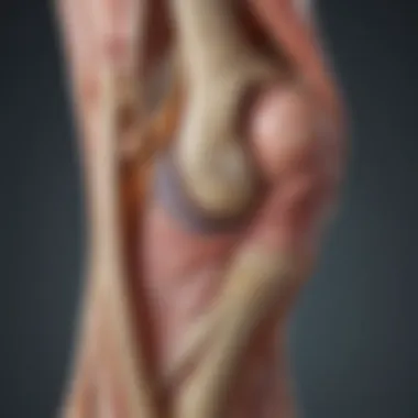
The posterior tibial tendon originates from the posterior tibial muscle, located in the deep posterior compartment of the leg. It travels down the back of the leg, passing behind the medial malleolus, and then inserts into multiple tarsal bones as well as the bases of the second, third, and fourth metatarsals. The anatomical design of this tendon allows it to provide support and stability to the arch of the foot, helping in the transition of weight during walking and running.
One vital aspect is the vascular supply. The posterior tibial tendon has a limited blood supply, especially at certain segments, which can complicate healing after an injury. This anatomical consideration plays a significant role when planning treatment options.
- Key anatomical features include:
- Origin: Posterior tibial muscle
- Pathway: Travels behind the medial malleolus
- Insertions: Tarsal bones and metatarsal bases
Function of the Posterior Tibial Tendon
The primary function of the posterior tibial tendon is to support the foot's arch and assist in foot inversion. When the foot begins to move, this tendon helps to control its positioning, allowing for proper gait and balance. It also plays a significant role in the push-off phase of walking.
In addition to arch support, it contributes to the foot's overall biomechanics. The tendon must be strong enough to withstand the forces exerted during various activities like running, jumping, and walking.
A rupture or significant tear can lead to structural changes in the foot, often resulting in flatfoot deformity. This change can lead to pain and functional limitations in daily life. Each of these functional aspects shows how critical understanding this tendon is for both diagnosis and treatment.
In summary, the posterior tibial tendon is not just a supportive structure; it is vital for functional foot mechanics and overall mobility.
Understanding the posterior tibial tendon allows for a better grasp of the implications associated with its injury. It sets the groundwork for accurate diagnosis, effective treatment options, and the preventive measures that can be taken to maintain ankle and foot health.
Etiology of Posterior Tibial Tendon Tears
Understanding the etiology of posterior tibial tendon tears is crucial for developing effective treatment strategies. This section emphasizes the underlying causes of such tears, encompassing both intrinsic and extrinsic factors. Allergies to these causes can inform prevention tactics and enhance recovery outcomes. By identifying risk elements and mechanisms leading to damage, clinicians can tailor interventions to meet individual patient needs.
Intrinsic Factors
Intrinsic factors refer to the internal conditions of the body that may predispose individuals to posterior tibial tendon tears.
- Age: Aging can lead to degenerative changes in the tendon structure. As people age, tendon elasticity decreases. This makes tendons more susceptible to injury during physical activities.
- Metabolic disorders: Conditions like diabetes can affect tendon health. Poor blood circulation may impair the body's ability to repair its tissues, increasing the likelihood of tears.
- Genetics: Certain genetic predispositions might worsen tendon conditions. Some individuals may have inherently weaker tendon structures. This weakness puts them at greater risk during high-impact activities.
- Foot mechanics: Biomechanical issues, such as flat feet or overpronation, place extra stress on the posterior tibial tendon. These altered mechanics can lead to increased tendon strain and subsequent injury.
Each of these intrinsic factors contribute to an individual's risk profile. Understanding them aids in proactive treatment and rehabilitation methods.
Extrinsic Factors
Extrinsic factors are external influences that impact the integrity of the posterior tibial tendon. Recognizing these can help in the assessment and management of potential injuries.
- Activity level: High levels of physical activity, particularly repetitive forceful motions, increase the chance of tendon injuries. Athletes or individuals engaged in certain sports may be at higher risk.
- Footwear choices: Inappropriate footwear can contribute to tendon stress. Shoes that do not provide adequate support can alter the natural mechanics of the foot. This results in higher stresses on the posterior tibial tendon.
- Environmental factors: Surface conditions, such as trails or uneven ground, influence the risk of tendon injury. Running or walking on hard surfaces can lead to increased tendon strain over time.
- Injury history: Previous injuries to the ankle or foot can increase the risk of developing posterior tibial tendon tears. Scar tissue or altered biomechanics post-injury may contribute to further damage.
Understanding extrinsic factors can lead to improved prevention strategies. Knowledge of how these factors interact with intrinsic factors provides a comprehensive view of injury development.
A thorough grasp of both intrinsic and extrinsic factors equips healthcare providers with the insights necessary to mitigate risks and enhance recovery for patients suffering from posterior tibial tendon tears.
Recognizing Symptoms of a Tear
Understanding how to recognize the symptoms of a posterior tibial tendon tear is critical for effective diagnosis and management of the condition. Early identification can lead to prompt intervention, which is essential for preserving foot functionality. The symptoms can vary widely among individuals and are often influenced by the extent of the injury. Notably, recognizing these signs can provide insights into the severity of the condition and guide treatment strategies.
Common Signs
The common signs of a posterior tibial tendon tear include:
- Pain: This is typically localized to the inside of the ankle, where the posterior tibial tendon runs. Pain may be sharp or a dull ache.
- Swelling: Patients often notice swelling around the ankle and foot area, which can indicate inflammation.
- Instability: Individuals may experience a sense of instability in the foot or ankle. This might manifest as difficulty walking or a sensation of "giving way" during movement.
- Flattening of the Arch: Over time, a tear may lead to a progressive flattening of the foot's arch, making it noticeable.
- Limited Mobility: Restrictions in movement, especially during activities requiring ankle mobility, may arise.
These signs, when observed early on, can facilitate timely evaluation by a healthcare provider. Prompt attention can prevent further deterioration and support more favorable recovery outcomes.
Assessment of Pain Levels
Assessing pain levels is a fundamental aspect of diagnosing a posterior tibial tendon tear. Pain assessment can be subjectively influenced by individual pain thresholds but remains a crucial indicator of injury severity. Clinicians often utilize various scales, such as the Numeric Pain Rating Scale, to quantify the pain experienced by the patient. Some factors to consider during the assessment include:
- Intensity of Pain: Rating the severity as mild, moderate, or severe.
- Radiation of Pain: Understanding if pain radiates to other areas, such as the heel or up the leg.
- Duration of Pain: Clarifying if the pain is constant or intermittent.
- Impact on Activities: Evaluating how pain affects daily activities and mobility.
Pain assessment provides critical data that informs succeeding diagnostic and treatment approaches.
In summary, recognizing the symptoms of a posterior tibial tendon tear and assessing pain levels are vital components in managing this condition. The insights gained through careful observation and evaluation can significantly influence treatment strategies, ultimately enhancing care outcomes.
Diagnostic Approaches
Understanding the diagnostic approaches to posterior tibial tendon tears is crucial for effective treatment. Early and accurate diagnosis can significantly influence the management plan and overall outcomes for patients. The diagnosis involves a combination of physical examination techniques and imaging studies. These methods help to confirm the presence of a tear, assess its severity, and identify any associated injuries.


The benefits of employing a comprehensive approach to diagnosis include improved patient outcomes and optimized treatment strategies. For instance, accurate physical assessment can reveal not just the location of pain but also the extent of functional impairment. Likewise, imaging studies provide necessary visual confirmation that complements the physical exam findings.
Physical Examination Techniques
Physical examination is one of the first steps in diagnosing posterior tibial tendon tears. The clinician typically starts with a thorough patient history to understand the onset of symptoms and any prior injuries. The physical exam often includes:
- Inspection: The foot and ankle area are examined for swelling, discoloration, or deformity.
- Palpation: The clinician uses their hands to feel for tenderness along the posterior tibial tendon.
- Functional Tests: Tests like the single-leg heel rise help assess the strength and functionality of the tendon. These tests can indicate whether there is significant weakness suggesting a tear.
Imaging Studies
Imaging studies play a key role in confirming the diagnosis of a posterior tibial tendon tear. They provide detailed images that help clinicians visualize the tendon and surrounding structures. Common imaging modalities include:
Ultrasound
Ultrasound is a widely used imaging technique for assessing soft tissue injuries. One significant aspect of ultrasound is its ability to provide real-time imaging, allowing the clinician to observe the tendon during movement. This feature makes it a valuable tool in diagnosing tendinopathy or tears. The key characteristic of ultrasound is its non-invasive nature and the lack of ionizing radiation. The advantages of ultrasound include its cost-effectiveness and accessibility. However, its effectiveness can be limited by the operator's experience and the patient's body habitus.
Magnetic Resonance Imaging
Magnetic resonance imaging (MRI) is another critical imaging modality for diagnosing posterior tibial tendon tears. MRI is particularly useful due to its high contrast resolution, which allows excellent visualization of soft tissues. The unique feature of MRI is that it provides detailed images of both the tendon and underlying cartilage, ligaments, and bones. MRI is often favored for its ability to provide comprehensive information without radiation exposure. However, it can be more expensive and may not be as accessible in certain clinical settings.
X-rays
X-rays are often the first imaging technique employed in cases of suspected tendon injury. They can reveal bone fractures or alignment issues, which might be contributing to the patient's symptoms. The key characteristic of X-rays is their ability to quickly visualize bone structures. This makes X-rays beneficial for identifying any underlying osseous pathology. However, X-rays do not show soft tissue details like tendons, thus limiting their diagnostic capacity for tendinous injuries.
In summary, while each imaging study has its strengths and weaknesses, a combination of physical examination and imaging studies maximizes diagnostic accuracy for posterior tibial tendon tears. This comprehensive approach aids in informing treatment strategies tailored to individual patient needs.
Conservative Management Strategies
Conservative management strategies play a crucial role in the treatment of posterior tibial tendon tears. These methods offer a non-invasive approach aimed at reducing pain, restoring function, and preventing further deterioration. Such strategies are preferred initially, as they allow the body to heal naturally while minimizing surgical risks. Understanding the key components of these strategies is essential for effective patient care.
RICE Method
The RICE method is a foundational approach in managing acute soft tissue injuries, including posterior tibial tendon tears. RICE stands for Rest, Ice, Compression, and Elevation.
- Rest: It is vital to avoid activities that put stress on the affected tendon. This includes walking or running, which can exacerbate the injury.
- Ice: Application of ice packs to the injuried area reduces swelling and alleviates pain. Ice should be applied for about 15-20 minutes every few hours in the first couple of days post-injury.
- Compression: Using compression wraps or bandages helps limit swelling. Proper application is important so as not to hinder blood circulation.
- Elevation: Keeping the affected foot elevated above the level of the heart promotes fluid drainage and reduces edema.
The RICE method, when applied correctly, can effectively manage symptoms in the acute phase, leading to improved outcomes.
Physical Therapy Interventions
Physical therapy is often essential in the conservative management of posterior tibial tendon tears. A qualified physical therapist can customize a program tailored to individual patient needs.
- Stretching Exercises: These help maintain flexibility in the calf and foot muscles, which can be beneficial in alleviating stress on the tendon.
- Strengthening Exercises: Gradually strengthening the muscles around the ankle enhances stability and support. This process is crucial for recovery and preventing future injuries.
- Manual Therapy: Techniques such as massage can improve circulation and relieve tension in the surrounding tissues.
Adherence to a structured physical therapy program can facilitate the healing process and restore function more effectively than relying solely on rest.
Orthotic Devices
Orthotic devices can provide additional support for those suffering from posterior tibial tendon tears. They aim to redistribute pressure and enhance foot biomechanics.
- Arch Supports: These help in maintaining the natural arch of the foot, which can alleviate strain on the tendon.
- Custom Footwear: Custom shoes can correct structural deformities and provide necessary support, allowing for safer mobility.
- Heel Lifts: These may assist in reducing the tension on the posterior tibial tendon by altering the foot's angle when walking or running.
Orthotic devices should be considered an integral part of the conservative management strategy, particularly for patients with ongoing symptoms.
For many patients, conservative management strategies can substantially improve their quality of life without the need for surgery. Their significance cannot be overstated, as they often lay the foundation for successful recovery.
Surgical Treatment Options
Surgical intervention becomes crucial when conservative measures fail to adequately address the symptoms and functional limitations associated with posterior tibial tendon tears. As the condition can lead to significant impairment in mobility and quality of life, understanding when surgery is appropriate is essential. Surgery for posterior tibial tendon injuries may offer restorative benefits, potentially allowing patients to regain their prior levels of activity and alleviate pain more effectively than non-operative treatments.
Indications for Surgery
Surgery is typically recommended for individuals who exhibit persistent symptoms despite rigorous conservative management, such as physical therapy and orthotic interventions. Key indications for surgical intervention may include:
- Inability to walk properly: If a patient struggles with daily activities.
- Significant pain: Unresponsive to medications or therapy.
- Progressive deformity: Such as worsening flatfoot or instability in the arch of the foot.
- Tendon rupture: Complete tears that fail to heal correctly require surgical correction.
Deciding when to proceed with surgery takes careful consideration of patient factors, including age, activity level, and overall health. The orthopedic surgeon will evaluate these elements before making a recommendation.
Types of Surgical Procedures
The surgical options for treating posterior tibial tendon tears can be diverse, each tailored to the specific needs of the patient. Here are some common procedures that are performed:
Tenosynovectomy
Tenosynovectomy involves cleaning out the sheath surrounding the tendon where inflammation is present. This procedure can help reduce pain and restore function by removing tissue that obstructs movement.
Key Characteristic: Tenosynovectomy focuses on the debridement of the tendon sheath, which may help promote healing in cases where inflammation leads to pain.
Benefits: This procedure is minimally invasive and often has fewer complications. It can lead to quicker recovery while preserving the tendon’s integrity.
Disadvantages: It may not fully restore function in cases of significant tendon damage or rupture, necessitating further procedures.
Tendon Repair
Tendon repair aims to restore the integrity of the torn tendon itself. This procedure involves suturing the ends of the tendon together.
Key Characteristic: A primary focus is on repairing the tendon to restore its original position and function.
Benefits: Tendon repair has the potential to completely restore function in many patients, leading to improved outcomes in motion and stability.
Disadvantages: The success of tendon repair can vary; it depends heavily on the degree of initial injury and the patient's adherence to post-operative rehabilitation plans.


Flexible Flatfoot Reconstruction
Flexible flatfoot reconstruction is a more comprehensive surgical procedure targeting both the tendon and the structural deformities of the foot. It involves restoring the arch by addressing the alignment and integrity of surrounding ligaments and tendons.
Key Characteristic: This procedure not only focuses on the tendon but corrects the biomechanical issues contributing to flatfoot deformities.
Benefits: Patients often experience a significant restoration of function. This procedure can result in improved foot mechanics and stability.
Disadvantages: It is a more complex surgery that may require a longer recovery time and carries a higher risk of complications compared to simpler procedures.
"Surgical options for posterior tibial tendon tears are tailored to individual patient needs and conditions, emphasizing restoration of function and relief of pain."
In summary, surgical treatment for posterior tibial tendon tear offers several options. The choice of procedure depends on the extent of the injury and the enhancement of the overall function of the foot. Each option comes with its unique advantages and challenges, necessitating thorough discussions between the patient and their surgical team.
Post-Surgical Rehabilitation
Post-surgical rehabilitation is a critical component in the recovery process for patients who have undergone surgery for posterior tibial tendon tears. It plays a vital role in restoring function, strength, and mobility. Rehabilitation not only enhances physical recovery but also contributes significantly to psychological well-being. Patients often face challenges post-surgery, including pain management and mobility limitations. Hence, structured rehabilitation protocols are essential to guide patients through these tough phases.
Effective rehabilitation programs typically begin shortly after surgery to reduce stiffness and promote blood circulation. These programs focus on various elements such as range-of-motion exercises and gradual loading of the affected tendon. It is crucial to personalize rehabilitation to fit each patient's specific needs and recovery timelines. This tailored approach ensures optimal outcomes, fostering motivation and adherence to the rehabilitation protocols.
Early Stage Rehabilitation
The early stage of rehabilitation begins immediately following the surgical procedure. The main objectives at this stage are to minimize swelling, manage pain, and prevent complications. Initial treatment may involve the use of ice and elevation. After a few days, physical therapy often commences with passive range-of-motion exercises.
During this phase, patients should be closely monitored for any signs of complications. Early rehabilitation not only helps to maintain the flexibility of surrounding tissues but also lays a foundation for more strenuous activities later on.
Common activities in early rehabilitation include:
- Isometric exercises to enhance muscle activation without stressing the tendon.
- Gentle stretching to improve flexibility around the ankle and foot.
- Use of assistive devices like crutches to aid mobility while protecting the surgical site.
Progressive Strengthening Exercises
As healing progresses, usually within weeks, the focus shifts to strengthening exercises. The goal is to regain strength and stability for functional activities. Progressive strengthening exercises involve controlled and graded loads to gradually increase the intensity of workouts.
These exercises typically start with closed-chain activities, where the foot remains on the ground. This method helps in retraining the posterior tibial tendon without putting excessive strain on it. Strengthening programs can include:
- Heel raises to target calf muscles, promoting strength in the entire lower leg.
- Balance exercises to improve proprioception and stability, essential for preventing future injuries.
- Resistance training that progresses systematically in intensity and complexity.
Incorporating these exercises not only restores muscle function but also enhances endurance in daily activities. Adherence to a well-structured progressive program can make a substantial difference to the patient's recovery trajectory.
"The pathway to recovery is often paved with consistent effort and gradual progress, leading to a restored quality of life."
In summary, post-surgical rehabilitation for posterior tibial tendon tears is integral to the overall treatment plan. It helps in managing the initial postoperative phase and empowers patients to regain their strength and mobility effectively.
Complications and Considerations
Understanding the complications and considerations related to posterior tibial tendon tears is crucial for both patients and healthcare providers. The risks associated with surgical interventions can greatly influence treatment decisions and long-term outcomes. A thorough analysis of potential complications helps patients make informed choices and prepares them for what to expect post-surgery.
Possible Surgical Risks
Surgery is often necessary in cases of severe tendon tears that fail to respond to conservative treatments. However, like any surgical procedure, there are inherent risks involved. Common surgical risks associated with posterior tibial tendon repair include:
- Infection: As with any surgical procedure, there is a risk of infection at the incision site. Proper preoperative preparation and postoperative care can help mitigate this risk.
- Blood Clots: The risk of thromboembolism increases during and after surgery, particularly in patients with limited mobility. Using compression devices and anticoagulant medications may reduce this risk.
- Nerve Damage: Surgical manipulation near nerves can sometimes lead to temporary or permanent nerve injury, resulting in numbness or weakness in the foot or ankle.
- Re-tear of the Tendon: While surgery aims to repair the tendon, there is always a chance of re-tearing during the healing process or due to excessive strain in the years after surgery.
"Informed patients who understand the risks can engage in open discussions with their healthcare providers, allowing for tailored treatment plans that suit their individual needs."
Long-Term Outcomes
The long-term outcomes of surgery for a posterior tibial tendon tear can vary widely among patients. Several factors influence the efficacy of surgical interventions, including:
- Timing of Surgery: Early intervention typically leads to better outcomes, as chronic tears may result in greater muscle and tendon degeneration.
- Post-Surgical Rehabilitation: The rehabilitation process following surgery is critical. Patients who adhere strictly to physical therapy and prescribed rehabilitation protocols tend to recover more effectively than those who do not.
- Age and Health Status: Younger patients or those with fewer comorbidities may experience faster recovery and better long-term function compared to older or less healthy individuals.
- Lifestyle Factors: Ongoing engagement in appropriate strengthening and flexibility exercises post-recovery can significantly improve outcomes and reduce recurrence risk.
Alternative and Adjunctive Therapies
Alternative and adjunctive therapies provide additional options for individuals recovering from a posterior tibial tendon tear. While conventional treatments such as surgery and physical therapy are essential, these complementary methods can enhance recovery, manage pain, and improve overall outcomes. They often focus on harnessing the body's natural healing processes, potentially avoiding more invasive tactics.
Integrating alternative therapies can lead to better patient outcomes. Each method has its unique benefits, and understanding these can help in making informed decisions about treatment. It is crucial, however, to consider the evidence supporting each therapy and consult with healthcare providers to ensure a safe and effective treatment plan.
Platelet-Rich Plasma Injections
Platelet-rich plasma (PRP) injections have gained attention in recent years as a promising alternative treatment. PRP involves drawing a small amount of the patient's blood, processing it to concentrate the platelets, and then injecting this solution into the site of the injury. The key advantage of PRP is its ability to accelerate healing through the growth factors found in platelets.
Benefits of PRP injections include:
- Promotion of Tissue Repair: Increased concentration of platelets helps foster a healing environment.
- Reduced Inflammation: PRP can help lower inflammation, thereby decreasing pain levels.
- Quick Recovery: Many patients experience a shorter recovery period when using PRP as part of their treatment plan.
Considerations regarding PRP therapy include its suitability for individual cases, potential side effects, and varying results based on specific conditions. Physicians often recommend PRP when patients have not responded well to standard treatments.
Stem Cell Therapy
Stem cell therapy is another cutting-edge approach gaining traction in treating tendon injuries. The premise is to utilize stem cells, which have the potential to differentiate into various cell types, promoting repair and regeneration within the damaged tendon. This therapy can be particularly valuable for those with chronic conditions.
The benefits of stem cell therapy include:
- Tissue Regeneration: By introducing stem cells, the body may experience enhanced regeneration of damaged tissues.
- Reduction in Scar Tissue Formation: This therapy can help minimize the formation of scar tissue, which can obstruct normal tendon function.
- Longer Lasting Results: Some studies suggest that stem cell therapy may produce longer-lasting improvements in function and pain relief compared to more traditional methods.
However, it is important to approach stem cell therapy with caution. Ongoing research is necessary to fully understand its efficacy and safety for specific conditions, particularly in the context of posterior tibial tendon injuries. Coordination with an experienced healthcare professional is vital for proper evaluation and implementation.
The exploration of alternative and adjunctive therapies can significantly shape the rehabilitation landscape for patients dealing with posterior tibial tendon tears. Adopting these methods may enhance recovery and provide hope for improved mobility and quality of life.
Preventive Measures


The significance of preventive measures in managing posterior tibial tendon tears cannot be overstated. Effective preventive strategies can considerably reduce the risk of injury and enhance overall foot health. Since these tendon tears often arise from a combination of intrinsic and extrinsic factors, addressing both aspects can contribute to a more resilient musculoskeletal system. Understanding and implementing preventive techniques helps in avoiding potential tears that may limit mobility and function.
Adopting a pragmatic approach involves a mix of exercises, proper footwear utilization, and awareness of body mechanics. These methods not only fortify the posterior tibial tendon but also prepare the foot for various physical activities. By emphasizing prevention, patients and practitioners can focus on maintaining optimal function and reducing the burden of treatment later down the line.
Strengthening and Flexibility Exercises
Strengthening and flexibility exercises play a vital role in the prevention of posterior tibial tendon tears. These activities enhance the resilience of the tendinous tissue while simultaneously improving the flexibility of associated muscles and joints.
A few effective exercises that can be incorporated into a routine include:
- Calf Raises: This exercise targets the calf muscles, which support the posterior tibial tendon during movements.
- Ankle Inversion and Eversion: These strengthen the muscles around the ankle and improve the tendon's overall stability.
- Toe Taps: A simple yet effective way to engage the foot's intrinsic muscles, promoting better arch support.
- Stretching the Achilles Tendon: Flexibility in this area reduces strain on the posterior tibial tendon, decreasing the risk of injury.
Incorporating these exercises regularly can lead to significant improvements in strength and flexibility. Furthermore, it encourages better biomechanical alignment, which is crucial for foot health.
Footwear Considerations
Choosing the right footwear is essential for preventing posterior tibial tendon injuries. Appropriate shoes can provide support and stability while accommodating the unique needs of the foot. Styles that lack adequate arch support or cushioning may exacerbate existing issues or lead to new injuries.
When selecting footwear, consider the following attributes:
- Arch Support: Shoes with proper arch support can help maintain alignment and reduce unnecessary stress on the posterior tibial tendon.
- Cushioning: Ample cushioning absorbs shock and reduces impact during activities, making a significant difference in comfort.
- Stability Features: Look for shoes designed with features that promote stability, particularly for those with overpronation.
- Fit: Ensure shoes fit correctly, allowing enough room for toes without excessive tightness.
Maintaining a regular evaluation of footwear is advisable, as shoe wear can impact support and comfort over time. Furthermore, investing in orthotic inserts may also provide additional support where necessary.
"The right footwear not only protects your feet but also supports your overall musculoskeletal health."
Continually adapting preventive measures, such as strengthening exercises and appropriate footwear, empowers individuals to maintain their foot health and minimize the risk of posterior tibial tendon tears.
Role of Patient Education
Patient education plays a crucial role in the management of posterior tibial tendon tears. Understanding the condition is the first step towards effective treatment and rehabilitation. It empowers patients to make informed decisions about their health. When patients comprehend the mechanics of their injuries, they become more engaged in their recovery process. They also better appreciate the necessity of following prescribed treatment plans.
Educating patients involves sharing detailed information about the anatomy and function of the posterior tibial tendon. When people learn how this tendon contributes to their foot's stability and movement, it enhances their awareness of the impact of a tear. They begin to recognize potential warning signs and symptoms, which can lead to earlier diagnosis and treatment. This is vital because early intervention is often linked to improved outcomes in tendon repair.
Moreover, education about conservative and surgical options ensures that patients have realistic expectations about their recovery. They need to know what each treatment entails, including potential benefits and risks. This not only fosters trust between patient and healthcare provider but also encourages adherence to treatment plans.
"An informed patient is a powerful ally in the treatment process."
Understanding the Condition
In order to adhere to treatment plans effectively, patients must grasp the implications of a posterior tibial tendon tear. The tendon itself supports the arch of the foot, playing a pivotal role in locomotion. Injuries can stem from various sources, including overuse, trauma, or underlying conditions such as diabetes and arthritis. Understanding these elements helps patients connect their experiences with general causes of the injury.
Additionally, understanding symptoms and their severity is essential. Patients often report pain, swelling, and sometimes, changes in foot positioning or difficulty while walking. Recognizing these symptoms can prompt patients to seek prompt medical attention, which is a critical factor in successful treatment. Educational resources can include pamphlets, reputable websites, and educational sessions led by healthcare professionals.
Importance of Adherence to Treatment Plans
Following treatment recommendations is vital for recovery. Patients who adhere to therapy and activity modifications tend to experience better results than those who do not. When patients understand the rationale behind their treatment regimen, they are more likely to meet the requirements for rehabilitation.
In the case of conservative management, it may involve a combination of rest, ice, compression, elevation, and physical therapy. If surgery is deemed necessary, adherence extends to postoperative rehabilitation plans. Understanding the stages of recovery can motivate patients to stay committed to their exercises and attend follow-up appointments.
Furthermore, ongoing support from healthcare providers throughout the treatment process can greatly increase adherence levels. Regularly scheduled check-ups offer patients the opportunity to address any concerns and adjust their treatment as necessary. Encouragement and understanding from providers can facilitate a more positive attitude towards recovery, emphasizing the patient's role in achieving lasting results.
Overall, patient education contributes significantly to effective treatment of posterior tibial tendon tears. An informed patient leads to improved outcomes, adherence, and ultimately, a better quality of life.
End
In this article, the conclusion serves as a critical synthesis of the extensive information regarding posterior tibial tendon tears. Understanding the treatment approaches discussed is vital for several reasons. First, it summarizes the comprehensive strategies available for managing, rehabilitating, and recovering from this common yet debilitating condition.
The emphasis on both conservative and surgical treatment methods reveals the complexity of care required. Conservative management, through techniques such as the RICE method and physical therapy, can often lead to significant improvements. However, recognizing when surgical intervention is necessary can make the difference between chronic pain and restored mobility. The balance of these options highlights the personalized nature of care.
Additionally, the role of patient education cannot be overstated. Patients equipped with knowledge about their condition and treatment options tend to adhere better to treatment plans. This ultimately enhances recovery outcomes, emphasizing that informed individuals are better positioned for success.
The article also outlines the considerations for rehabilitation post-surgery, which stresses the importance of following a structured recovery plan. Long-term outcomes depend not only on the initial treatment but also on the commitment to rehabilitation. As we acknowledge the evolving nature of orthopedic practices, staying updated with latest innovations will help practitioners provide the best care.
"A comprehensive understanding of posterior tibial tendon tears enables practitioners to make informed decisions, fostering the best possible outcomes for their patients."
This comprehensive overview not only assists practitioners but also offers students and researchers valuable insights into the multifaceted nature of treatment for posterior tibial tendon tears.
Future Directions in Research
Research into posterior tibial tendon tears is essential for advancements in treatment methodologies and improved patient outcomes. Understanding the nuances of this condition not only facilitates better treatment plans but also aids in the overall comprehension of foot biomechanics. Future research endeavors should focus on several specific elements that can lead to enhanced therapeutic interventions and beneficial clinical practices.
The exploration of new treatment options continues to be a top priority. Current methods, while effective, have limitations that can affect patient recovery. A comprehensive overview of innovative treatment methods—ranging from biologic therapies like platelet-rich plasma to novel surgical techniques—could pave the way for more efficient care. Researchers must assess how these methods impact pain relief and functional recovery in patients suffering from posterior tibial tendon tears.
Furthermore, understanding the long-term efficacy of existing treatments is vital. As patients undergo various modalities, it is imperative to establish which interventions yield sustainable outcomes. This involves systematically tracking recovery patterns and any associated complications over extended periods. Evaluating long-term data can help pinpoint which treatments provide the most lasting benefits, ultimately contributing to tailored treatment strategies for individual patients.
Continued research not only enhances treatment options but also educates practitioners on optimal patient management strategies.
The significance of patient education on the management of posterior tibial tendon tears must also be examined. Investigating effective ways to communicate treatment plans to patients can influence adherence to rehabilitation guidelines and overall satisfaction with care.
In summary, future research on posterior tibial tendon tears should prioritize innovative treatment methods, long-term efficacy assessments, and patient education strategies. These areas hold the potential for substantial advancements in orthopedics and improved quality of life for patients affected by this debilitating condition.
Innovations in Treatment Methods
Innovative treatment methods for posterior tibial tendon tears are crucial in improving recovery outcomes and functional mobility. One area of focus is the development of biologic therapies, such as platelet-rich plasma and stem cell injections, which have gained attention in recent years. These therapies aim to utilize the body's healing capabilities to promote tissue repair and regeneration.
Another innovation involves enhanced surgical techniques. Minimally invasive procedures, such as arthroscopic tendon repair, are being refined to reduce recovery times and surgical complications. Surgeons are also exploring the role of tendon scaffolds that may support healing in cases of severe injury, providing a framework for tissue regeneration that aligns with the body's natural recovery processes.
Conventional methods often underestimate the importance of individualized treatment plans. Personalized approaches that consider each patient's unique biomechanics and lifestyle are becoming increasingly important. Through precise evaluations, medical professionals can better tailor interventions, such as orthotic support or custom rehabilitation protocols, to align with the patient's specific needs and activities.
Research on Long-Term Efficacy
Assessing the long-term efficacy of treatments for posterior tibial tendon tears is vital in determining the best practices for managing this condition. Longitudinal studies focusing on patient outcomes can yield valuable insights into how different therapies perform over time.
Evaluating the durability of surgical repairs and conservative management are critical areas for future research. Metrics such as pain levels, functional capacity, and overall satisfaction should be meticulously recorded. Researchers need to follow up with patients at regular intervals post-treatment to gauge efficacy and adapt current practices accordingly.
Moreover, understanding how demographic factors influence treatment success can provide a holistic view of recovery from posterior tibial tendon tears. By identifying variables such as age, activity levels, and previous injuries, researchers can better predict outcomes and refine treatment algorithms.
In summary, detailed research into long-term efficacy is necessary to establish the most effective, sustainable treatment methods for individuals with posterior tibial tendon tears. By focusing on this area, healthcare professionals can ensure that patients receive the highest quality care tailored to their needs.


